
Artificial intelligence (AI) is becoming increasingly important in dentistry and healthcare. AI uses advanced computer technology to replicate human-like critical thinking, decision-making, and intelligent behavior. So, artificial intelligence applications in dentistry mainly include decision-making for prognosis, diagnosis, and therapy.
In healthcare, AI exists in virtual and physical forms. Virtual AI includes tools for medication dosages, diagnosis, scheduling, drug interactions, electronic health records, and imaging. Physical AI involves rehabilitation, telepresence, robotic surgery assistance, and robots for elderly care.
AI operates in two key phases: training and testing. During training, the model learns from existing data, like patient records or datasets, to determine its parameters. In testing, it uses these parameters to make predictions on new data. The AI process follows a basic structure of Input, Processing, and Output.
In dentistry, input data can include voice commands, medical records, or images like radiographs.
Neural networks process this data to analyze clinical cues, perform cephalometric studies, or detect lesions. The output might be a diagnosis, prognosis, treatment plan, or disease prediction. AI can simulate outcomes, predict treatments, and connect data to CAD/CAM systems, enhancing precision and efficiency.
While AI has the potential to revolutionize medicine and dentistry by solving clinical problems and simplifying workflows, its applications in dentistry are not yet routine. This blog focuses on examples of the application of AI in dentistry, especially in endodontics. It explores AI’s role in diagnosis, clinical decision-making, and predicting successful therapy.
.
Quick Summary
This summary covers the key applications of AI in endodontics:
- Disease Diagnosis: AI can identify conditions with high precision, even at an early stage.
- Treatment Planning: It helps create effective and personalized treatment plans.
- Image Analysis: AI analyzes radiographs and other images to detect small changes or abnormalities.
- Prognosis Prediction: It predicts treatment outcomes and the success of retreatment procedures.
- Root Canal Anatomy: AI studies complex root canal structures for better treatment accuracy.
- Fracture Detection: It identifies root fractures that might not be visible to the human eye.
- Lesion Identification: AI detects periapical lesions and tracks their progression.
.
Have more time? Keep reading for an in-depth look and comprehensive insights.
.
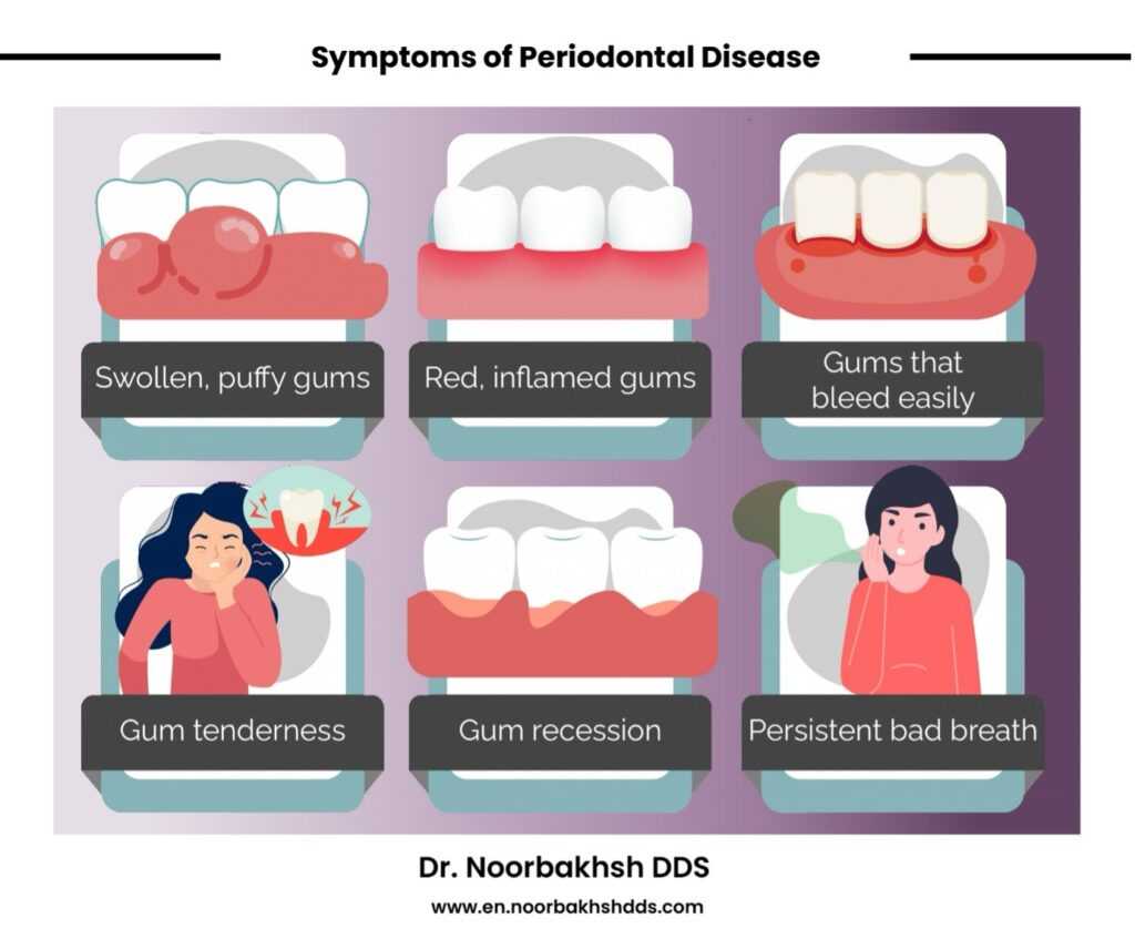
Periapical Lesion Detection
Diagnosing and planning treatment for teeth with periapical lesions can be difficult, as about 75% of radiolucent jaw lesions are caused by apical periodontitis (a type of inflammation at the tip of a tooth root). Early detection improves treatment success, prevents the spread of infection to surrounding tissues, and reduces complications.
Clinicians often use two common 2D diagnostic methods: IOPA (intraoral periapical radiographs, which are small X-ray images focused on the tooth and surrounding bone) and OPG (orthopantomograms, which are panoramic X-rays showing all teeth and the jaw). In these methods, periapical lesions appear as dark spots (radiolucencies) on the X-rays. However, since 2D images flatten 3D structures, they can be unreliable. CBCT (cone-beam computed tomography) imaging, which provides 3D views, offers more precise detection of periapical lesions by showing their exact location and size. However, CBCT is less effective for diagnosing lesions in teeth with root fillings.
AI significantly advances the detection of periapical lesions by analyzing features like radiolucency and alveolar bone resorption (loss of bone around the tooth socket). AI models now detect and measure bone loss with precision. Deep learning techniques identify severely affected teeth and predict those unlikely to be saved. Advanced models classify the severity of periapical lesions and detect them with accuracy comparable to experienced oral surgeons. Additionally, AI methods differentiate granulomas, which can heal after treatment, from cysts using CBCT images. Learn more about CT scans and CBCT imaging in our blog on digital dentistry.

Image Sources: First Image, Second Image, Third Image (From Left to Right)
.
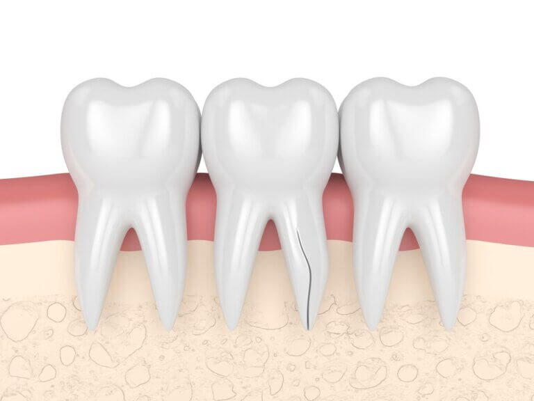
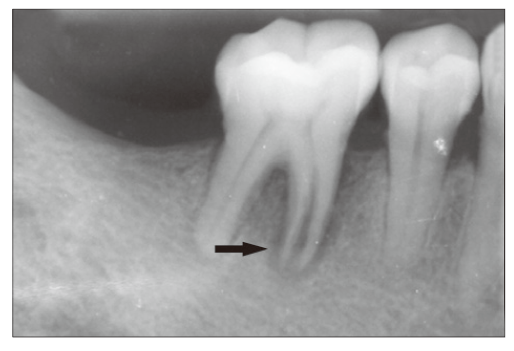
Root Fracture Detection
Vertical root fractures (VRFs), which account for 2% to 5% of crown/root fractures, often require root resection or tooth extraction. Diagnosing VRFs can be challenging, and unclear diagnoses may lead to unnecessary surgeries or extractions. Cone-beam computed tomography (CBCT), a 3D imaging technique, and traditional radiographs are used to identify VRFs, but radiography often has low sensitivity and may miss fractures. CBCT imaging offers better accuracy, sensitivity, and specificity compared to 2D radiographs.
AI have been effective in identifying VRFs on panoramic radiographs. Neural networks have been trained using CBCT and periapical radiographs to detect fractures in both intact and root-filled teeth and have been used successfully to detect fractures in high-resolution CBCT images.

Image Sources: Image 1&2, Image 3, Image 4
.
Determination of Working Length (WL)
Accurate determination of working length (WL) is critical for successful root canal treatment. WL is the distance from the coronal reference point to the root canal’s apical endpoint. Common methods for assessing WL include radiography (X-ray imaging), digital tactile sense (a manual method relying on feel), electronic apex locators (devices that measure electrical resistance to locate the canal endpoint), patient reactions to a paper or file point placed in the canal, and cone-beam computed tomography (CBCT), a 3D imaging method. Dentists most often rely on radiography and electronic apex locators for routine WL assessment. Clear radiographic images are essential for interpreting the anatomy of the root canal system accurately. However, factors like image quality or interpretation errors can lead to misdiagnosis.
To improve consistency, AI is used. AI can provide a second opinion by accurately locating the radiographic apical foramen (the canal’s endpoint). Research has shown that AI, when tested in a clinical setting using a human cadaver model, produced WL measurements that matched real root lengths, and in some cases even outperformed endodontists.

Image Source
.
Understanding Root and Root Canal Structure
Understanding root structure and root canal systems is essential for successful nonsurgical root canal therapy. Dentists use X-rays and a 3D imaging technique called CBCT (a type of scan that shows the whole tooth in 3D). CBCT is more accurate than X-ray for determining root canal structures, but it is not recommended for routine use due to radiation risks.
AI has brought significant advancements in this area, as well. For example, AI algorithms can differentiate complex root structures, for example in certain back teeth (the distal roots of mandibular first molars) quickly and accurately. Additionally, AI can analyze CBCT segmentations as accurately as human operators, but much faster. These advancements demonstrate the potential of AI in improving root canal treatment precision and efficiency.
See this AI-powered CBCT imaging demo as an example of how AI is already enhancing CBCT imaging.
.
Decision on Tooth Retreatment
AI can help dentists decide if a root canal retreatment (fixing a tooth again) will work or not. The AI system looks at similar cases from the past to figure out the risks and benefits of doing the retreatment. This system uses statistics from other patients to make predictions about whether the treatment will be successful.
This system can accurately predict retreatment outcomes based on the available data. But, how well it works depends on the quality and diversity of information it is given for analysis. If the data is not good or comes from only a few examples, the predictions might not be as accurate. To make it better, researchers need to include more examples from different types of cases so it can learn and make smarter decisions.
.
Stem Cell Survival Testing
In a study, scientists used AI to check if stem cells (special cells from the inside of a tooth) could stay alive after certain treatments. These treatments were tested to see how the cells would react to germs that cause infections.
To do this, they added a substance from bacteria to the cells to make them act like they were fighting an infection. The smart system then guessed whether the cells would survive, and it was able to make accurate predictions. This helps doctors know how well these treatments might work in real-life situations.
.
In Dental Education
AI has made a significant development in learning process for dental students. With the help of AI system, dental students can engage with scenarios that simulate clinical work on patients without the need to work with real patients. This makes learning safer and helps students avoid mistakes on live patients.
These AI programs give feedback to students, showing them how to improve their skills. Students can check their work and compare it to how it should look, helping them learn faster. Studies show that students get better at their work more quickly using these systems than with older training tools.

AR and AI in dental education. Image Source
.
Augmented and Virtual Reality in Dentistry
.
For Patient Management
AI-powered virtual dental assistants perform various tasks in dental clinics with high precision and fewer errors compared to human staff. They can book appointments, record detailed medical and dental histories, manage insurance, and assist dentists in diagnosis and treatment planning. These systems also alert dentists to patient habits, such as tobacco or alcohol use, and other relevant medical history, by creating a comprehensive virtual database of patient information.
Virtual dental assistants are also valuable for online emergency health consultations, offering timely support to patients when needed. Please read our blog on how teledentistry works for more information.
.
For Diagnosis, Treatment, and Prognosis
AI is highly beneficial in diagnosing and treating oral diseases, including detecting and classifying suspicious mucosal changes that may indicate premalignant or malignant conditions. It can identify even tiny changes at the pixel level that might be missed by the human eye. AI systems can also analyze genetic data to identify a population’s risk for oral cancer. In determining a dental prognosis, AI models are valuable tools. They help evaluate a tooth’s long-term health and function by integrating detailed treatment strategies, ensuring better decision-making and outcomes.
.
In Oral and Maxillofacial Surgery
AI has changed oral surgery with the development of robotic systems that replicate human motion and decision-making. These systems are used for procedures such as dental implants, tumor removal, foreign object removal, biopsies (checking tissues for disease), and TMJ surgery (temporomandibular or jaw joint). This robotic -assisted surgeries show significantly better accuracy compared to freehand methods, even when done by skilled surgeons.
Studies found no performance difference between experienced surgeons and trainees using these systems. Robotic surgery also reduces operation time, increases accuracy, and ensures safer procedures around delicate structures. These procedures are semi-automated, meaning that skilled surgeons still guide these robotic systems to make sure everything is done correctly, but the AI system is making surgery better and easier for both patients and surgeons.

Perceptive dental robotic system (Photo credit: Perceptive)
.
In Prosthetic Dentistry
A special tool called RaPid considers things like face size, shape, background, and what the patient likes and helps dentists make the most suitable prosthesis. This system connects databases, knowledge-based systems, and computer-aided design (CAD), learn from patterns and get better over time in designing perfect prosthesis that fit better, work well, and look great. These systems design and produce inlays, onlays, crowns, and bridges with high accuracy, replacing the traditional method of casting prostheses.
.
In Orthodontics
AI is transforming orthodontics by helping every step including diagnosis, treatment planning, and monitoring. Radiographs and images from intraoral scanners help for diagnosis and planning, and there’s no need for multiple lab procedures or patient impressions.
These AI-assisted tools often provide results more accurate than human perception. Using 3D scans and virtual models, aligners can be 3D printed to match a personalized treatment plan. AI algorithms decide how each tooth should move, how much pressure to apply, and where to apply it for the best results. The result is shorten treatment time, minimal errors and easier appointments.
See this demo as an example of how AI can enhance orthodontics.
.
In Forensic Odontology
In forensic odontology, AI is widely used to determine a person’s age and gender, analyze bite marks, and predict the shape of the jawbone.

Image Source
.
Innovations in Dental Technology for Future
Voice-command dental chairs
In dental offices, new technology includes voice-activated chairs that dentists can control without touching them. These chairs might soon be able to check vital signs, monitor anxiety levels, track weight, and measure how long procedures take, all while keeping the patient comfortable and alerting dentists to any problems.
.
Bioprinting
Another exciting advancement is bioprinting, a method that creates living tissues and organs layer by layer. In the future, this could help rebuild damaged or lost soft and hard tissues like gums or jawbones, due to disease or injury.
.
Conclusion
Artificial intelligence (AI) is widely used in endodontics. Research shows that neural networks perform as well as dental experts, with higher accuracy and precision in some cases. In certain studies, AI has even outperformed specialists. AI is especially helpful for beginners and non-specialists by offering expert-level guidance.
Here’s the key benefits of Dental Application of AI:
- Diagnostic Support: Deep learning provides assistance to general dentists.
- Automation: AI speeds up clinical processes, like automatically completing electronic dental records by identifying and numbering teeth.
- Productivity Boost: Automated systems increase efficiency.
- Secondary Views: AI offers a second opinion to improve diagnostic precision.
AI should be seen as a tool to assist dentists, not replace them. It helps by integrating patient information and improving professional relationships. While AI excels at processing structured data and analyzing large datasets, it cannot think like the human brain. It struggles with making complex decisions in unclear clinical situations.
Good patient-dentist communication depends on interpreting nonverbal cues, such as hopes, fears, and expectations. While there are debates about adding empathy to AI, these intuitive communication skills remain unique to humans and cannot be fully replicated by machines.
Reference
This blog offers a simplified and audience-friendly adaptation of the research article:
Agrawal P, Nikhade P. Artificial Intelligence in Dentistry: Past, Present, and Future. Cureus. 2022 Jul 28;14(7):e27405. doi: 10.7759/cureus.27405. PMID: 36046326; PMCID: PMC9418762.
Available in: https://pubmed.ncbi.nlm.nih.gov/36046326/


2 Responses
Keep up the excellent piece of work, I read few blog posts on this web site and I conceive that your website is really interesting and contains sets of excellent info .
Thank you for your wonderful feedback! I’m delighted to hear you enjoy the blog and find the information useful.